Surgical Techniques Algorithm in Morton's neuroma
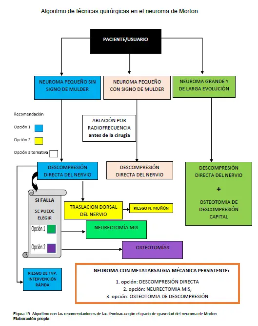
When the patient presents with a small Morton’s neuroma or without Mulder’s signs, the first option recommended is Direct decompression of the nerve by releasing the deep transverse intermetatarsal ligament.
As a second option we would have the dorsal translation of the nerve.
When the patient presents with a symptomatic small Morton’s neuroma and a positive Mulder’s sign, the first option would be the direct decompression.
As an alternative to surgery, we can perform the radiofrequency ablation Using it before surgery, if there is no improvement then option 1 (direct decompression) could be performed.
If the neuroma fails direct decompression, the first option may be to choose the neurectomy by CMI and as a second option the decompression osteotomies.
In large and long-standing neuromas, it is recommended combination of techniques, carry out release of the deep transverse ligament next to decompression osteotomies.
If the neuroma presents with persistent mechanical metatarsalgia, the first option would be direct decompressionthe second option CMI neurectomy and the third option decompression osteotomy.
If there is a risk of stump neuroma, the best option is dorsal translation of the nerve And in cases of patients at high risk of DVT where you need to minimize the intervention time, the chosen technique is… direct nerve decompression.
The ultrasound-guided endoscopic nerve decompressionIt is a good option for resection of the deep transverse intermetatarsal ligament, but several months must be waited to observe improvement in pain, compared to the ultrasound-guided nerve decompressionDirect ultrasound-guided nerve decompression has better short-term results, which is why this technique was chosen in the logarithm.

NM is a neuropathy of the common digital nerves, originating from the medial plantar nerve or medial (internal) branch. Nerve entrapment usually occurs at the anastomosis of the medial plantar nerve and the lateral plantar nerve. (5)
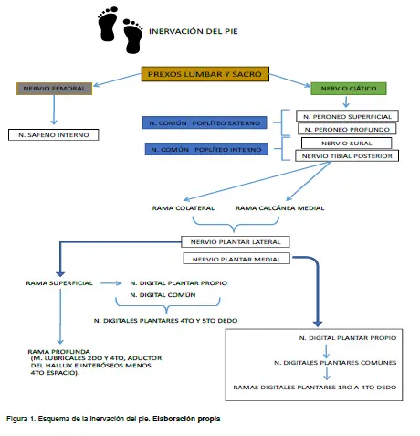
Figure 1 schematically shows the nerve innervation in the foot.
Figure 2 shows the plantar innervation of the posterior tibial nerve with its lateral branch (lateral plantar nerve), medial branch (medial plantar nerve) and its variants.
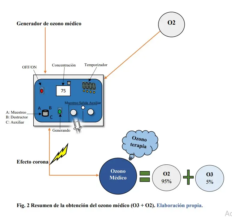
The ozone therapy It is the therapeutic application of medical ozone, which is a gaseous mixture composed of oxygen (not less than 95%) and ozone¹ (not exceeding 5%), by means of an electric generator that obtains ozone from pure oxygen, passing through a high voltage gradient.
We set out to analyze the indications, effectiveness and safety, as well as evaluate the advantages and disadvantages of ozone treatment.
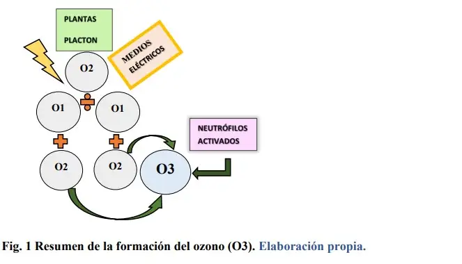
Figure 1 shows the different mechanisms for obtaining ozone (O3), as well as its formation.
Figure 2 shows the mechanism for obtaining medical ozone used in ozone therapy.